
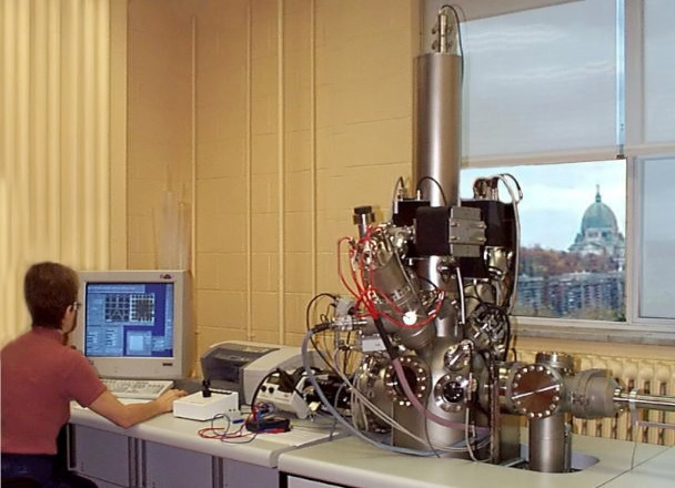
 This modern, powerful, and sensitive technique is used to analyze the elemental and chemical composition of materials surface including conductive and insulating samples such as minerals and organics (polymers, biological materials, etc.).
The process is based on the irradiation of a material surface by an energetic primary ion beam that sputters the surface at energies between 1 and 30 kev. This causes the emission of secondary particles (atoms, ions, molecules) which are analyzed via conventional mass spectroscopic techniques.
Information provided:
- Depth profiles up to many microns can be obtained by analyzing and monitoring the ions removed as a function of time.
- The mass, position, and intensity of each secondary ion is recorded at each pixel to provide information with a special resolution along with the intensity of each species.
- The distribution of chemical species can be defined with a 1 µm spatial resolution, and the mass is defined within two milli-atomic mass units or better.
This technique is widely used for investigating the chemistry of solid surfaces. Materials that are either conductive or insulating can be analyzed. The sample is irradiated with photons from a soft X-ray source with a well defined energy. This causes core electrons with lower binding energy (Eb) than the energy of the incident photons to be ejected from the atoms. The kinetic energy (Ek) of the emitted electrons is given by: Ek = hv - Eb - Φ, where hv is the energy of the X-ray photon, and Φ is the work function, the energy needed for the electron to free itself from the surface. Since the kinetic energy of the emitted photoelectrons is unique for different elements, both elemental and chemical state information for all elements of the periodic table, except helium and hydrogen, are provided by this technique.
Information provided:
- Atomic surface composition.
- Chemical environment.
- Oxidation number.
- Depth profile.
- Overlayer thickness.
- Plasmons, shake-ups.
- Inelastic background.
- Fermi level.
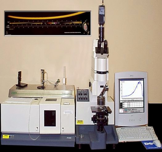
This rapid, precise and convenient spectral technique is a non-destructive method that provides information about the types of bonds present in a material and can often enable us to identify the exact material being used. Infrared (IR) radiation is used to excite the vibrational modes of a molecule in the gas phase or absorbed on a surface. The transmitted or reflected IR spectrum can be analyzed using a spectrometer. Absorption bands appear as peaks in the spectrum. A considerable improvement of sensitivity is achieved by use the Fourier Transform method. We have two systems:
- FTIR- ATR (Attenuated Total Reflection) used to enhance the surface sensitivity.
- FTIR- PAS ( Photoacoustic ) used to analyze material in two modes : Step-scan and Rapid-scan, and can be used for depth profiles.
Information provided:
- Molecular bonds.
- Molecular identification.
- Surface-chemical reactions.
- Branching.
- Orientation of absorbed molecules.
Advantages:
- Can determine protein secondary structure in liquids or solids.
- Can analyze small samples of different shapes (fibers, particles), copolymers, films, semi-solids, gels and liquids.
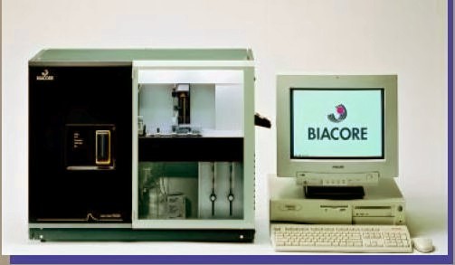
The Biacore System is an optical biosensor, based on the surface plasmon resonance (SPR) technique, which measures the change in the angle of surface plasmon resonance due to the surface binding of large molecules. This allows the real time monitoring of specific interactions between surface-bound ligands and macromolecules in many biologically relevant cases.
The method has a growing number of applications, such as:
- studying kinetics of antibody-antigen interactions.
- monitoring solid phase gene assembly.
- following enzymatic activity on DNA and hybridization.
- studying interactions between surface-bound ligands and RNA.
In the Biacore apparatus, analyte flows over immobilized ligand, and an SPR signal, which depends on changes in the mass concentration at the sensor-surface layer, is detected in real time.
The method provides detailed information about equilibrium and/or kinetic binding constants.
These sensors represent one of the foremost types of direct, label-free observation of biomolecular interaction. Surface plasmons are longitudinal oscillations of free charges on the surface of the metal film that can be excited under specific conditions of total internal reflection of plane-polarized light. Under these conditions, the wave vector of the evanescent wave is equal to the wave vector of the surface of plasmon and a fraction of the incident light is absorbed, thereby diminishing the intensity of the reflected light. This occurs for a specific angle of incidence (θ°), at a given wavelength (λ), and is a function of the dielectric properties of the sample medium and, therefore, its refractive index.
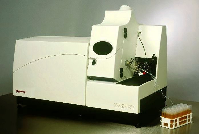
ICP-MS is a method for trace element analysis. It is capable of multi-element analysis for most elements in the mass range of lithium to uranium. The liquid sample is introduced into the plasma. In the case of solid sample, a suitable preparation method is required (dissolution, extraction, etc.).
ICP-MS couples the excitation source of inductively coupled argon plasma with a quadrupole mass spectrometer. Ions produced in the plasma pass through the orifice into an interface region that is maintained at 1 Torr. The ions are sampled through the next orifice in a second cone to form an ion beam. A series of ion optics further forms the ion beam and rejects photons also generated by the argon plasma. The quadrupole mass spectrometer separates the ions according to their mass-to-charge ratio, permitting only a single, specified mass to pass through at any given time. The ions are detected by channel electron multiplier.
Advantages:
- A rapid multi-elemental technique that can provide meaningful data on at least 75 elements within minutes.
- A dynamic range that is linear over 5 to 6 orders of magnitude.
- Determination of elemental isotopic concentrations and ratio.
- Spectra are relatively simple and easy to interpret.
Applications:
- Alloys.
- Polymers.
- Ceramics.
- Biological materials.
- Pharmaceuticals.
- High-purity mineral acids.
- Organic solvents.

The LV-SEM is a powerful technique that has greatly improved the possibility of analyzing a wide variety of unique specimens in a nondestructive manner.
This tool incorporates the functions of a High Vacuum SEM and Low Vacuum SEM in a single instrument and is coupled with an Energy Dispersive X-ray Spectrometer (EDS).
Many advantages make this method attractive:
- The possibility of working with uncoated samples which can be preserved for further use.
- Dehydratation in a graded ethanol series is not necessary.
- The possibility of varying vacuum, which allows the observation of biological products and gels without provoking an evaporation of the liquids or a decrease of the charge effect.
- Many organic and inorganic, conductive and insulting materials can be easily observed and identified.
- It offers the option of observing samples at different temperatures (-50 à 80°C).
- Energy Dispersive X-ray Spectrometer (EDS) detects all characteristic X-ray emitted from a specimen simultaneously.
- LV-SEM / EDS permits a specimen containing many types of elements to be analyzed in a short time.
- Full quantitative and qualitative analysis procedures can be combined with X-ray digital maps, area and line element distribution using the LV-SEM-EDS integration features.
Low Vacuum SEM is, therefore, a simple, time-saving and reproducible method that is applicable to various specimens.
This system, unique in Quebec, is used with the LV-SEM for the preparation of biological samples. It has many advantages:
- It carries out fine cutting through soft tissues, like muscles or bone marrow, hard materials, like dental enamel, without damaging any of the materials or disrupting the interface between the different substances.
- It polishes flat surfaces with low forces that minimizes distortion or other structural impact to the sample.
- LThe surface quality of the cut is significantly improved over that of line cutting.
- The time required to make the cut is reduced.
- Less force is translated directly to the sample and less heat is generated at the cutting surface.
- The amount of cutting debris that is introduced to the surface of the cut is reduced.
 This device is a non-contact 3D capture system, capable of acquiring dense geometric data from complex surfaces.
A beam of light coming from a single source (a laser, a lamp, etc.) is split into multiple light beams using two or more flat mirrors.
These beams are then combined to create an interference pattern.
The desired result is to find alternating bands of light called fringes.
Fringes are bright where the beams are constructively adding together and dark where they are canceling each other out.
This tool presents a wide range of applications. All kinds of materials in various states of processing and even translucent objects can be measured with an accuracy of a few microns or even nanometers without contact.
It enables us to carry out many types of measurements:
- Measuring individual parts of shape acquisition and inspection purpose.
- Measuring even ceramic, silicon, human skin, rubber, etc.
- Roughness measurement.
We use this technique especially to determine the surface topography of a biomaterial which allows us to assess the roughness and porosity.

This instrument is used to measure the contact angle between a liquid and a biomaterial surface in order to determine the wettability and the surface free energy, which is an indication of the hydrophobic/hydrophilic behavior of materials. The wettability is a very important aspect of many surfaces, especially in coating applications. Contact angle from 0°-30° are typical for a hydrophilic surface, whereas contact angle > 60° indicate a hydrophobic surface. Surfaces with extreme hydrophobicity are known to produce angles up to 150°.
Provided information:
- Uppermost surface information (~ 1nm).
- Surface tension (surface free energy).
- Hydrophilic / hydrophobic balance.
- Surface relaxations.
- Contact hysterisis.
- Roughness.
Applications:
- Study of adhesion.
- Wetting behavior.
- Bonding quality.
- Surface treatment and coating on biomaterials, fiber, polymer, semi-conductor wafer..
- Absorption studies.
This thermal analysis equipment is linked to a Mass Spectroscope and allows us to study the thermal profile and degradation behavior of a sample.
Differential scanning calorimeter (DSC) measurements:
The change in temperature and heat flow, associated with transitions in materials, as a function of time and temperature. Such measurements provide qualitative and quantitative information about physical and chemical changes that involve endothermic and exothermic processes, or changes in heat capacity.
Thermo-gravimetric analysis (TGA) measurements:
The mass of sample changes as a function of temperature. This information may be used for assessing compositions or to characterize phenomena such as evaporation and drying, decomposition, oxidation, and oxidative stability.
Atomic force microscopy is a technique for measuring surface topography. Uses for this technique, for studying protein adsorption, surface-induced thrombosis and cell-surface interactions, are well-known. It provides unique opportunities for visualizing surface-dependent cellular and molecular interactions in three dimensions, on a nanometer (nm) scale, and in aqueous environments. The principle implies a direct force measurement between a small tip (r ~ 50 nm) and a surface. The deflection of the cantilever (force-measuring spring, < 1N/m) is detected using a reflected laser beam. Normal (cantilever deflection) and lateral (cantilever torsion) forces can be detected using a four segment photodiode.
A computer can acquire a two dimensional representation of the topology of constant tip-surface interaction which can include Van der Waals forces, electrostatic interactions, specific interactions, and capillary forces.
Provided information:
- Topography.
- Tip-surface forces.
- Local mechanical properties.
- Nano-tribology (friction).

Flow cytometry is used to rapidly count and identify bacteria, cells, and other biological particles. The flow cytometer functions by passing cells through a laser beam in a continuous, single-file stream. Each cell scatters some of the laser light, and also emits fluorescent light excited by the laser. The cytometer measures a number of factors, based on scattering and emission, and uses these data to differentiate and count the types of cells in the mixture.
For flow-cytometric analysis, The XL system features the capability of:
- Analysis up to 4 colors of immunofluorescence.
- Throughput of up to100 samples per hour.
Other multi-color applications include:
- Multiparametric DNA analysis.
- Platelet studies.
- Reticulocyte enumeration.
- Cell biology/functional studies.
- Broad range of research applications.
This apparatus is widely used in cell biology, biochemistry and molecular biology to determine:
- Solution molecular weight, and to assess the degree of homogeneity.
- The stoichiometry and equilibrium constants for interacting macrosolutes.
- The overall hydrodynamic shape of a macrosolute in solution.
- Whether aggregates exist, and if it’s a reversible or an irreversible process.
Tow methods are used to provide data:
1. Sedimentation velocity that is performed at higher rotor speed and allows calculation of:
- Sedimentation coefficient.
- Diffusion coefficient.
- Effective mass.
- Shape information.
- Molecular weight.
2. Sedimentation equilibrium that performed at lower rotor speed and allows calculation of:
- molecular weight.
- homogeneity.
- association constant.
- aggregation states.
These systems are highly sensitive methods that allow us to study platelet aggregation.
Application fields are:
- Toxicology.
- Diagnostics of thrombocyte stage of homeostasis.
- Assessment of platelet vitality during blood transfusion.
- Diagnostics of congenital and acquired homeostatic disturbances.
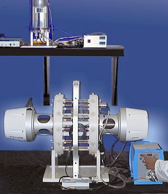
The SGT 9100 apparatus is one of the most efficient facilities for testing stent fatigue that use non-contact laser measurement system to measure simulated vessel compliances.
Natural rubber latex tubing is used to simulate human arteries and two electro-dynamic pump assemblies on either side of the saline filled latex tubing re-create the systolic and diastolic pressure within the simulated arteries. The pumps move axially towards and away from each other to displace the fluid. The latex tubing expands or constricts to keep the volume constant. It is this constriction and expansion that mimics the action of the human artery and applies the circumferential forces to the stent.
A laser measurement system monitors and records the tubing?s compliance (diameter as a function of pressure).
- The machine is capable of maintaining a pre-set vessel compliance and electronically ensures a file with various test output and input test parameters.
- With a maximal capacity of 12 stents, this machine is designed to conduct accelerated fatigue tests in physiological-like conditions at variable frequencies (40-60 Hz) for stents with diameters varying between 5 and 10 mm.
- The duration of fatigue test depends on the design of the stents. The test time can vary from 3 to 5 months for ~ 400 million cycles and depends on the test fatigue cycle determined during the initial testing of the stents.
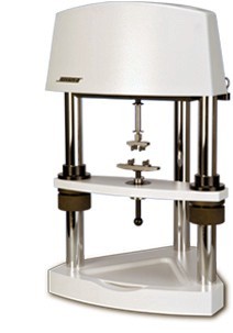
The MTS-Bionix system is a servo-hydraulics traction machine. It provides flexibility when performing lower force dynamic and static testing and allows unparalleled capabilities for performing a wide variety of tests on materials, biomaterials, components, etc.
Characterization of biomaterial properties:
- Yield and ultimate strength.
- Creep and viscoelastic characteristics.
- Fatigue characteristics.
- Fracture toughness and fracture mechanics.
- Wear characteristics.
- Coefficient of thermal expansion.
- Modulus of elasticity.
- Poisson’s ratio.
The ElectroForce 3200 test instrument provides precise fatigue testing of biomaterials and biological specimens within a closed environment. The ability to precisely load specimens and simulate in vivo biological conditions is needed for medical devices and tissue engineering. The instrument can be used to evaluate biomaterials, scaffolds, native tissue samples and tissue-engineered construct.
This equipment is necessary to draw biomedical grade nanocomposites fibers which are attracting attention in tissue engineering (artery, cartilage, muscles and nerve scaffolds) and will be developed for tissue reconstruction and repair. Such equipment is not available for research purposes in Canada. The equipment will be installed at IMI, for similar reasons and conditions as the aforementioned equipment.
 For our histological, SEM and ToF-SIMS studies of harder materials such as metallic, ceramic and nanofibre composite implants, we require specimen preparation equipment capable of cutting and grinding samples while preserving the interface between the implant and biological tissue in order to appreciate tissue ingrowth and integration. This sophisticated sectioning system is also needed to observe highly resolved topographical and compositional features of sectioned hard materials.
 TD System 3000 produces a very stable and thermally efficient flame that delivers up to 140,000 Btu/hr. (when used with propylene fuel).
This high energy level makes it possible to produce premium quality coatings while spraying at very high deposition rates..... up to 45lbs./hr.
 These systems extend the range of testing, providing an order of magnitude improvement in displacement measurement at low amplitudes.
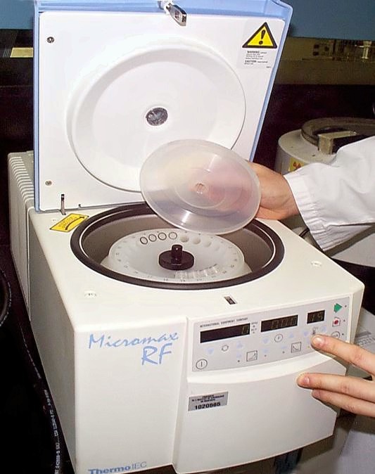
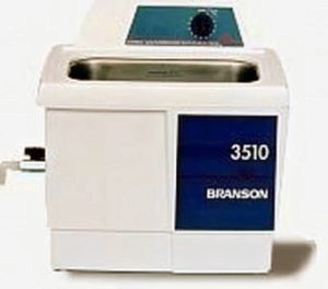 This 5.7L ultrasonic cleaner is new. The Branson ultrasonic cleaners remove blood, proteins and contaminants associated with sterilization of medical and dental instruments, and flux from electronics. They also deep clean to remove dirt, grease, waxes and oils used from switches, gears, and metal and plastic parts, and assemblies. The cleaning tank is recessed for fewer spills and drips. A durable, impact-resistant plastic enclosure withstands the toughest cleaning solutions and environments. The enclosure is lightweight for benchtop use. Tanks are rugged stainless steel. Sweep frequency ensures even cleaning throughout the tank. All units feature 40Hz industrial stacked transducers.
 Designed to operate in 37 C (98.6 F) incubator environment or on the lab bench for general purpose rotation. Features a compact, space-saving design and quiet, efficient operation. Ridged, silicon-rubber mixing platform lifts off easily for cleaning. Platform will accommodate various sizes of well plates, printed well slides, culture plates, flasks, beakers and vials.