Department of Electrical Engineering
Biomedical engineering Biomedical technology Life sciences research related to human health and disease Medical sciences Image and video processing Artificial intelligence Experimental methods and instrumentation Medical physics
Human Health
Modeling and Artificial Intelligence
Research interests and affiliations
My lab is developing advanced Magnetic Resonance Imaging (MRI) techniques for quantitative assessment of the brain and spinal cord structure. These developments include hardware (coils), MRI sequences (relaxometry, diffusion tensor imaging, magnetization transfer, functional MRI) and software (multimodal registration, segmentation, motion correction, distortion correction). There is a strong focus on translating research developments into clinics, aiming to improve the diagnosis and prognosis in patients suffering from neurological diseases and traumas.
The environment is highly multi-disciplinary. We interact with biomedical engineers, physicists, radiologists, neurologists and neurophysiologists. Plus, our lab collaborates with the Martinos Center for Biomedical Imaging at Harvard, therefore some projects will involve travelling to Boston. Projects include:
- Developing methods for spinal cord MRI in multiple sclerosis (collaboration with Harvard),
- Studying features of the brain using ultra-high field MRI (collaboration with Harvard),
- Development of deep learning methods for analysis of medical images (collaboration with Mila),
- Development of open source software for medical imaging applications,
- Building of MRI antenna
- Tier-2 Canada Research Chair in Quantitative Magnetic Resonance Imaging, Chairholder
- Functional Neuroimaging Unit (UNF), Co-director
- Laboratoire de Recherche en Neuroimagerie (NeuroPoly), Co-director
- Centre de recherche du CHU Sainte-Justine, Université de Montréal, Montreal, QC, Canada, Researcher
- Centre de recherche de l'Institut universitaire de gériatrie de Montréal (CRIUGM), Researcher
- Department of Neurosciences, Université de Montréal, Adjunct Professor
- Quebec Bio-Imaging Network (QBIN), Member
- Advanced Research Centre in Microwaves and Space Electronics (POLY-GRAMES), Member
- Institut de génie biomédical, Member
- Institute for Data Valorization (IVADO), Member
- Mila - Quebec AI Institute, Montreal, QC, Canada, Associate member
- 1900 BIOMEDICAL ENGINEERING
- 1901 Biomedical technology
- 6400 LIFE SCIENCES RESEARCH RELATED TO HUMAN HEALTH AND DISEASE
- 9000 MEDICAL SCIENCES
- 2708 Image and video processing
- 2605 Pattern analysis and machine intelligence
- 2603 Computer vision
- 2705 Software and development
- 3115 Medical physics
Publications


Biography
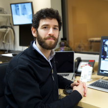
Prof. Cohen-Adad is an MR physicist and software developer with over 15 years of experience in advanced MRI methods for quantitative assessment of the brain and spinal cord structure and function. He is an associate professor at Polytechnique Montreal, adjunct professor of neuroscience at the University of Montreal, associate director of the Neuroimaging Functional Unit (Univ. Montreal), member of Mila (Univ. Montreal) and he holds the Canada Research Chair in Quantitative Magnetic Resonance Imaging. Along with his colleagues Prof. Nikola Stikov, Prof. Alonso-Ortiz and Prof. De Leener, he is co-directing the NeuroPoly Lab (www.neuro.polymtl.ca), which includes about 30 graduate students and research associates. Prof. Cohen-Adad's research is highly cited (Google Scholar). As a leader in the field, he organized multiple workshops at international conferences (https://spinalcordmri.org/workshops.html). He is a frequent guest lecturer on advanced MRI methods and he regularly serves as consultant for various companies (e.g. Biospective Inc., NeuroRx, IMEKA) and academic (Harvard, U. Toronto, UCL, UCSF, etc.) for setting up MRI acquisition and image processing protocols.
Do you want to join the lab? See: job opportunities
Teaching
- GBM6125 : Bases du génie biomédical
- GBM6904 : Séminaires de génie biomédical
- GBM8378 : Principes d'imagerie biomédicale
Supervision at Polytechnique
COMPLETED
-
Ph.D. Thesis (6)
- Enguix Chiral, V. (2022). Neonatal Resting-State Functional Magnetic Resonance Imaging: Optimization of Data Acquisition and Democratization of Data Preprocessing [Ph.D. thesis, Polytechnique Montréal].
- Mangeat, G. (2021). Development of in-Vivo Histology with Quantitative Magnetic Resonance Imaging to Resolve Fine Neurodegenerative Features [Ph.D. thesis, Polytechnique Montréal].
- Topfer, R. (2021). Local Coils and Prospective Shimming for MRI of the Spinal Cord [Ph.D. thesis, Polytechnique Montréal].
- De Leener, B. (2017). Development of an MRI Template and Analysis Pipeline for the Spinal Cord and Application in Patients with Spinal Cord Injury [Ph.D. thesis, École Polytechnique de Montréal].
- Duval, T. (2017). Quantification de la microstructure de la moelle épinière humaine par IRM et application chez des patients avec sclérose en plaques [Ph.D. thesis, École Polytechnique de Montréal].
- Lopez Rios, N. (2017). Development of a New Multi-Channel MRI Coil Optimized for Brain Studies in Human Neonates [Ph.D. thesis, École Polytechnique de Montréal].
- Enguix Chiral, V. (2022). Neonatal Resting-State Functional Magnetic Resonance Imaging: Optimization of Data Acquisition and Democratization of Data Preprocessing [Ph.D. thesis, Polytechnique Montréal].
-
Master's Thesis (21)
- Banerjee, R. (2025). Automated Segmentation of the Spinal Cord on Magnetic Resonance Echo Planar Images [Master's thesis, Polytechnique Montréal].
- Bouthillier, M. (2025). Outcomes Prediction in Acute Cervical Traumatic Spinal Cord Injury from Clinical and Preoperative Magnetic Resonance Imaging-Based Models: The Role of Imaging Biomarkers [Master's thesis, Polytechnique Montréal].
- Bréhéret, A. (2025). Impact of Through-Slice Gradient Optimization for Dynamic Slice-Wise Shimming in the Cervico-Thoracic Spinal Cord and the Brain [Master's thesis, Polytechnique Montréal].
- Bédard, S. (2024). Exploring the Impact of Anatomical References for Quantifying Spinal Cord Morphometrics Using Magnetic Resonance Imaging [Master's thesis, Polytechnique Montréal].
- Bourget, M.-H. (2022). Écosystème logiciel de segmentation de neurones sur des images microscopiques par apprentissage profond [Master's thesis, Polytechnique Montréal].
- Cereza, G. (2022). Development of an Innovative Solution Minimizing RF Field Inhomogeneity and Energy Deposition in Ultra-High Field MRI Applications [Master's thesis, Polytechnique Montréal].
- D'Astous, A. (2022). Shimming Toolbox: An Open-Source Software Package for Shimming the B0 and B1 Field in Magnetic Resonance Imaging [Master's thesis, Polytechnique Montréal].
- Lemay, A. (2022). Impact of Soft Segmentation Training on Medical Image Segmentation and Uncertainty Representation [Master's thesis, Polytechnique Montréal].
- Vincent, O. (2021). Impact of Rater Style on Deep Learning Segmentation in Medical Imaging [Master's thesis, Polytechnique Montréal].
- Rouhier, L. (2020). Prognosis for Degenerative Cervical Myelopathy: A Computer Learning Approach on the AOspine Database [Master's thesis, Polytechnique Montréal].
- Nami, H. (2019). Clustering of the White Matter Tracts in the Rat Spinal Cord Based on Quantitative Histology [Master's thesis, Polytechnique Montréal].
- Samuel Perone, C. (2019). Deep Learning Methods for MRI Spinal Cord Gray Matter Segmentation [Master's thesis, Polytechnique Montréal].
- Gros, C. (2018). Automatic Segmentation of Intramedullary Multiple Sclerosis Lesions [Master's thesis, École Polytechnique de Montréal].
- Zaimi, A. (2018). Automatic Axon and Myelin Segmentation of Microscopy Images and Morphometrics Extraction [Master's thesis, École Polytechnique de Montréal].
- Germain, G. (2017). Une antenne hybride RF/shimming pour l'IRM de la moelle épinière [Master's thesis, École Polytechnique de Montréal].
- Saliani, A. (2017). Construction d'un atlas de la microstructure de la matière blanche de la moelle épinière chez le rat à partir d'acquisitions histologiques [Master's thesis, École Polytechnique de Montréal].
- Vuong, M. T. (2017). Comparison of Myelin Imaging Techniques in Ex Vivo Spinal Cord [Master's thesis, École Polytechnique de Montréal].
- Lévy, S. (2016). Caractérisation de la microstructure des voies spinales humaines par IRM multiparamétrique [Master's thesis, École Polytechnique de Montréal].
- Mangeat, G. (2016). Study of Human Cortical Microstructure Using Magnetization Transfer and T2* Mapping with Application in Multiple Sclerosis [Master's thesis, École Polytechnique de Montréal].
- Foias, A. (2015). Design and Construction of a Highly Sensitive Coil for MRI of the Spinal Cord [Master's thesis, École Polytechnique de Montréal].
- De Leener, B. (2014). Segmentation automatique de la moelle épinière sur des images de résonance magnétique par propagation de modèles déformables [Master's thesis, École Polytechnique de Montréal].
- Banerjee, R. (2025). Automated Segmentation of the Spinal Cord on Magnetic Resonance Echo Planar Images [Master's thesis, Polytechnique Montréal].
News about Julien Cohen-Adad
Press review about Julien Cohen-Adad


