
Blogue
Giant leaps forward in spinal cord imaging
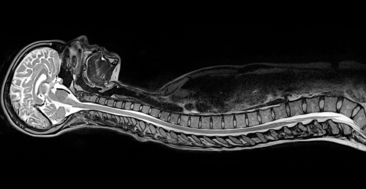
Thanks to a protocol developed by Professor Julien Cohen-Adad and his collaborators, it's now possible to use AI to read spinal cord MRI data. (Photo : NeuroPoly)
Not content with just one, two articles penned by Cohen-Adad and his team have just been published in the Nature group of journals. The research represents a contribution that could boost the development of treatments to combat neurodegenerative diseases, and spinal cord lesions. All this, thanks to a protocol that standardizes the collection of data obtained via magnetic resonance imaging (MRI).
Since its arrival in hospitals around the world during the 1980s and 1990s, MRI has given modern medicine giant leaps forward by making it possible to see what's happening inside patients' bodies without having to resort to surgery.
MRI is used to detect pathologies such as tumors anywhere in the body, especially in the brain and spinal cord, and also permit us to observe lesions associated with neurodegenerative diseases such as multiple sclerosis (MS) or amyotrophic lateral sclerosis (ALS).
Yet, there is a catch...
Until recently, spinal cord imaging data could only be used qualitatively - the lack of a reference point in the images made it impossible to calibrate what could be measured.
For example, it's difficult to follow the evolution of a patient's disease or to measure improvement in their health post-treatment, even if the images have been captured by the same device and the same technician only a few months apart.
“Neuroimaging devices must be configured for each use, you can't just press a button and make them work,” explains Professor Julien Cohen-Adad, from Polytechnique Montréal's Department of Electrical Engineering.
“Also, MRI applications have mainly been developed for the brain. For spinal cords, there was still a technology gap," adds Professor Cohen-Ada, who is also Co-director of the Neuro-Imaging Research Laboratory (NeuroPoly).
Note the use of the past tense here - how come? Well, that gap is well on its way to being closed thanks to the work of Professor Cohen-Adad and his collaborators.
Two impactful publications
 Julien Cohen-Adad (Photo : Polytechnique Montréal) Julien Cohen-Adad (Photo : Polytechnique Montréal) |
The research group has just published two impactful articles - with Professor Cohen-Adad as first author - in two of the Nature group of magazines, Nature Protocols and Scientific data.
The first article details the steps of a protocol that permits the “normalization” of spinal cord images taken by MRI, regardless of the device's manufacturer. The second article demonstrates the effectiveness of the protocol. The articles summarize a colossal project carried out over five years, and which involved 42 imaging centers across the globe, from America, to Asia, via Europe. In total, images from 260 individuals have made it possible to constitute an "open source" database.
“To develop new treatments, we first need to be able to see the pathology, but also to understand how it evolves. Now we have access to a quantitative spinal cord MRI database that will allow us to do this," notes Cohen-Adad.
This data will make it possible to standardize images using biomarkers, or by measuring the size of certain structures. It will also be used in laboratories like the Professor's for automated learning (AI) to improve patient health diagnostic and measurement tools.
"For example, we can follow the evolution of a patient's disease or see how they react to treatment," notes Professor Cohen-Adad, providing the example of patients suffering from partial spinal cord lesions who have been able to improve their condition after rigorous training on a treadmill.
The protocol developed by Cohen-Adad and his team will also accelerate the segmentation of spinal cord tumors (i.e. determining their stage) by making it possible to quantify tumor volume during each imaging session. It will also render possible the quantification of the speed with which neurodegenerative diseases such as MS or ALS progress. "The baseline diagnosis is made, among other things, by counting the number of spinal cord lesions," explains the NeuroPoly Co-director.
The tool will also prove invaluable for surgeons looking to determine whether a patient is operable or not. "We're working on spinal cord segmentation methods to help surgeons in their decision-making," adds Professor Cohen-Adad.
AI's contribution
 These seeming Christmas decorations are actually 3D prints of the brains of students in Professor Cohen-Adad's lab who volunteered to take an MRI test. (Photo : NeuroPoly)
All of this work is expected to accelerate with the contribution of Artificial Intelligence, and work carried out by members of the team supervised by the Polytechnique Montréal professor.
By standardizing spinal cord imaging data, the end result will be training algorithms - just like those we already use to detect various types of cancer elsewhere in the body.
“We're still developing methods. One of the big limitations that we’ve encountered so far was the quality of data obtained by various methods." This however is a problem that will disappear if the protocol proposed by the Professor and his international collaborators is adopted.
A glance at... MRI |
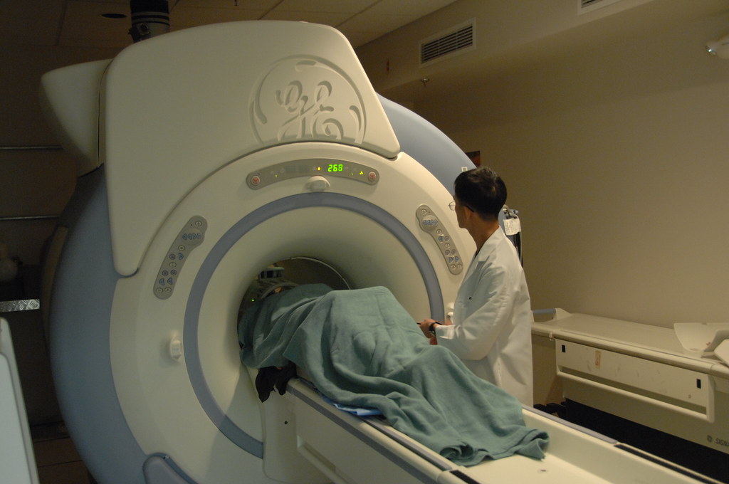 MRI machines use a powerful magnet coupled with radio waves to create 2D and 3D images of the interior of a human body. (Photo : National Institutes of Health (NIH), using licence CC PDM 1.0). MRI machines use a powerful magnet coupled with radio waves to create 2D and 3D images of the interior of a human body. (Photo : National Institutes of Health (NIH), using licence CC PDM 1.0).Unlike a CT scan or x-ray, MRI does not rely on radiography. Rather, it works using the behaviour of the tinest hydrogen atoms, placed in the center of a strong magnetic field. Hydrogen atoms are found in water molecules, in every organic molecule in the human body, and are found everywhere in different concentrations depending on the nature of the tissue. At the center of these atoms, a proton spins on itself, much like the Earth does - but on a whole different scale. Normally, a proton spins without a precise direction. This changes however, when those atoms are placed in a magnetic field like that of an MRI machine, which then magnetizes and aligns its rotation with the magnetic field, a bit like the needle of a compass placed in the same magnetic field. Then, radio waves emitted at regular intervals by the MRI machine come into play. These waves temporarily deflect the protons' rotation axis. When the radio signal disappears, the protons realign themselves according to the magnetic field and release a radio wave. This is the signal that the imaging device picks up. Once processed by computers, this data is used to reconstruct 2D or 3D images of the location targeted in the body by the device. Each tissue contains different concentrations of water - and therefore different concentrations of hydrogen atoms - and so location in the body is determined. |
En savoir plus
Professor Julien Cohen-Adad's expertise
NeuroPoly Lab's website
Nature Protocols article
Scientific data article
Department of Mechanical Engineering website


Comments
Commenter
* champs obligatoire