
Research project title
Quantitative imaging in MRI
Education level
Master or doctorate
Director/co-director
Director: Eva Alonso Ortiz
End of display
September 1, 2027
Areas of expertise
Mathematical biology and physiology
Unit(s) and department(s)
Department of Electrical Engineering
Detailed description
Description:
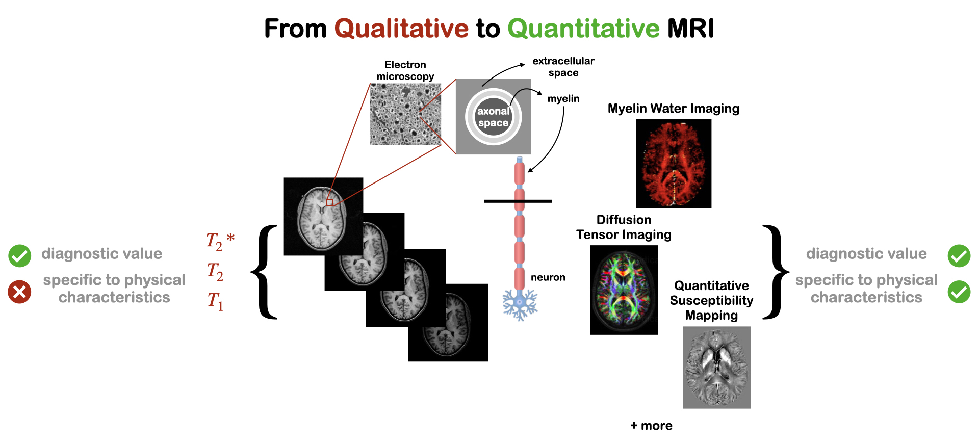
MR images have very high soft-tissue contrast and can convey macrostructural, microstructural, physiological and functional information. These types of images are commonly used in clinical settings (for diagnosis and treatment planning) and in research where MRI is used as an investigative tool (for ex: in neuroscience and neuropsychology). They are considered qualitative images. This means that the image contrast depends on intrinsic tissue properties, as well as specifics of the experiment. A consequence of that is that images cannot be compared across groups and changes in contrast are not specific to any particular aspect of the physiology. Quantitative MRI (qMRI) on the other hand, has the potential to deliver images that are specific to tissue characteristics and independent of the hardware and software used to acquire the images. The benefits of such images is that they allow researchers to investigate biological changes due to disease, aging, or normal development. By acquiring qMRI data in different populations, we can create databases of representative values in healthy and diseased groups. The efficacy of treatments for neurological diseases could then be evaluated by comparing subtle changes in qMRI measurements with the expected values for that population, allowing for greater diagnostic specificity and sensitivity, compared to what is achievable with qualitative MRI. qMRI can also eliminate problems of bias and interpretation, given that changes in images are no longer subjectively interpreted (by identifying abnormally bright, dark, small or large signal changes), but objectively measured.
Example projects: Develop a qMRI model that accounts for the magnetic susceptibility of iron and myelin in order to differentiate between them, study the effects of the spinal cord’s curvature on qMRI measures of myelin. All research will be carried out at the NeuroPoly lab (Polytechnique Montreal, www.neuro.polymtl.ca), at the Unité de Neuroimagerie Fonctionnelle (www.unf-montreal.ca), and at the Centre d’imagerie cérébrale McConnell (www.mcgill.ca/bic).
Contact: Eva Alonso Ortiz
Financing possibility
This project has available financing
