
Nouvelles
World first: a Polytechnique Montréal team pushes the boundaries of non-invasive brain imaging
A team at Polytechnique Montréal is advancing vascular imaging technology toward clinical use, potentially enabling early detection of neurodegenerative diseases. In a breakthrough that pushes back the limits of the technology, the group has, for the first time, imaged the brain’s smallest blood vessels—the capillaries—without directly accessing the brain. The results were published today in the prestigious journal Proceedings of the National Academy of Sciences (PNAS).
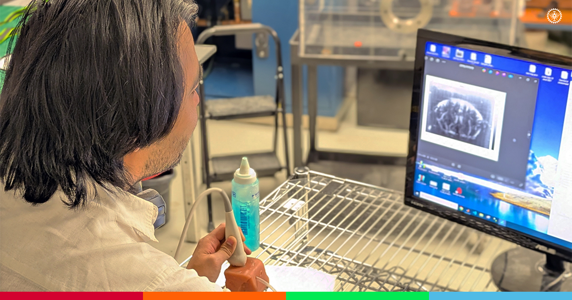
Stephen Lee, postdoctoral fellow, demonstrates the capabilities of the ultrasound probe to Jean Provost, Professor in the Department of Engineering Physics. (Photo : Martin Primeau)
Diagnosing brain disorders such as Alzheimer’s disease and vascular dementia remains one of the great challenges in modern medicine. These conditions often go undetected in the early stages, as does damage caused by some types of strokes, which are minor but can signal more serious future events.
Though the symptoms may not be immediately apparent, these conditions cause subtle changes in the brain. Blood flow in the capillaries is altered and, in some cases, red blood cells may stall. If the situation persists, it can be fatal to surrounding cells, starving them of oxygen and nutrients while preventing waste removal.
These changes are known to exist but are difficult to detect due to their microscopic scale, which evades current medical imaging tools. However, a promising new method—ultrasound localization microscopy (ULM)—could change that.
Pushing the boundaries of visibility with ultrasound
 Prof. Jean Provost (Photo : Polytechnique Montréal) |
Like conventional ultrasound, ULM uses a probe placed on the skin to show what’s happening inside the body. But instead of displaying the features of a foetus or the condition of the appendix, it maps the blood system by tracking tiny microbubbles as they move through the blood vessels. These microbubbles are already used in cardiology.
Despite its potential, ULM faces significant challenges in practice. A research team led by Polytechnique Montréal engineering physics professor Jean Provost has overcome some of these obstacles.
“The main limitation for ultrasound microscopy is the resolution of the images it produces,” explains Provost. “Capillaries are so small that they only let through one red blood cell or microbubble at a time—and these are already smaller than the resolution limit.”
Another challenge is time. Since microbubbles pass through capillaries infrequently, it takes several minutes of data collection to capture enough images of different microbubbles to fully map the capillary network, making the process difficult to implement.
When American postdoctoral researcher Stephen Lee joined the Polytechnique lab in 2022, he was aware of these issues and determined to find a solution.
“We knew the information required to image the capillaries was embedded in each scan in some way,” says Lee. “The challenge was figuring out how to extract it.”
He suggested a radically different approach: instead of reconstructing images based on all the microbubbles tracked over an extended period, it might be enough to track just one microbubble.
He was right.
a game changer
Video excerpt from the PNAS article showing how the “capillary reporters” (SCaRe) approach enables the visualization of individual capillaries. (Source : Stephen Lee)
To travel from an artery to a vein, a microbubble must pass through a capillary. Therefore, Lee reasoned, researchers tracking a single microbubble would observe a typical speed pattern—fast in arteries, slow in capillaries, then fast again in veins—and this U-shaped profile would serve as a functional fingerprint.
This led to the development of single capillary reporters (SCaRe), which are described in today’s PNAS article. Instead of treating a microbubble as a single point to be used to reconstruct a complete representation of a capillary, the researchers analyze its movement from image to image to track its trajectory and measure the changes in velocity. When a microbubble slows or stops briefly, it is passing through a capillary.
SCaRe not only images capillaries but also makes it possible to assess their condition by measuring microbubble transit time, a method the team demonstrated using a mouse model of neuroinflammation and validated with computer simulations.
“It is suspected that ‘stalling’—where blood flow temporarily stops in a capillary—may signal future problems,” Provost explains. “For the first time, we’ve visualized this using ultrasound microscopy.”
SCaRe doesn’t make ULM a ready-to-use clinical tool just yet. Scanning time is still long, data processing is complex and 3D imaging remains a goal. But demonstrating that it is possible to extract functional data from infrequent microbubble events is a game-changer. SCaRe could enable the use of ULM for early detection of neurodegenerative diseases without invasive procedures.
Provost’s team plans to collaborate with Montreal hospitals in the coming months to further develop this approach.
Learn more
“Assessing cerebral capillary function and stalling using single capillary reporters in ultrasound localization microscopy,” published on January 9, 2026 in PNAS
Jean Provost's faculty profile
Department of Engineering Physics website

